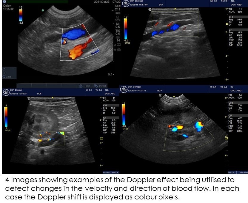
Ultrasound imaging in B-mode, color and spectral Doppler of the
4.6 (511) In stock
Download scientific diagram | Ultrasound imaging in B-mode, color and spectral Doppler of the abdominal organs of the agouti. (a) Ultrasonographic aspects of the urinary vesicle. Note the smooth and echogenic walls with a slight amount of sediment on the interior. (b,d) Color flow and B-mode renal morphology of the right and left kidneys, respectively, showing the usual echotexture and parenchymal echogenicity and preserved corticomedullary limit. (c,e) Pattern of flow of the renal artery, arcuate (arrowhead) and interlobar (arrow) arteries observed with color Doppler. The pulsed Doppler demonstrates well-defined systolic and diastolic peaks. from publication: Abdominal B-mode and Doppler ultrasonography of chemically restrained agouti (Dasyprocta prymnolopha Wagler, 1831) | Agoutis are small-sized wild animals whose body weight can reach up to 4kg, and are found throughout Brazil. They are considered important seed dispersers, especially for big trees and there are species that rely almost exclusively on these animals for their territorial | Doppler Ultrasonography, Doppler Ultrasound and Hemodynamics | ResearchGate, the professional network for scientists.

Francisco SOUSA, Laboratório de Fisiologia, PhD, Universidade Estadual do Piauí (UESPI), Teresina, Centro de Ciências da Saúde
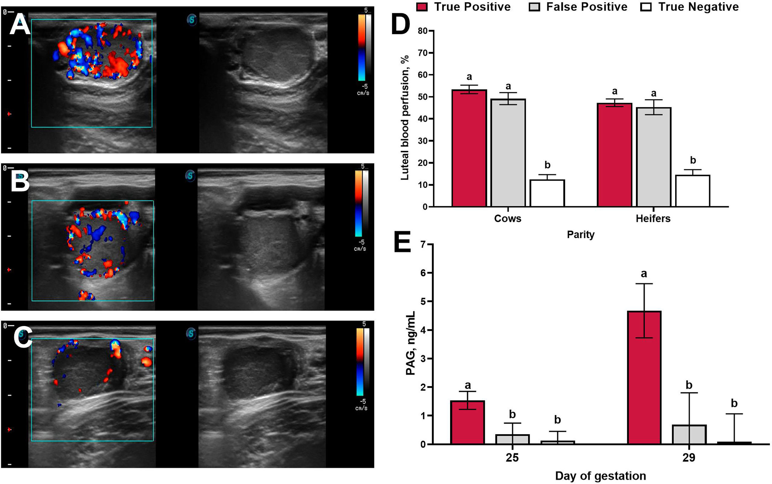
Frontiers Applied Use of Doppler Ultrasonography in Bovine Reproduction

Francisco SOUSA, Laboratório de Fisiologia, PhD, Universidade Estadual do Piauí (UESPI), Teresina, Centro de Ciências da Saúde

Francisco SOUSA, Laboratório de Fisiologia, PhD, Universidade Estadual do Piauí (UESPI), Teresina, Centro de Ciências da Saúde

Figure 1 from Manycore GPU Based High Performance Implementation of Ultrasound Color Doppler Imaging

Flávio ALVES, Laboratory Head, Ph.D, Universidade Federal do Piauí, Teresina, UFPI, Departamento de Morfofisiologia Veterinária
Macrophages in the liver and muscle of lean and obese mice.

Color Doppler Ultrasound Scnaner with Good 4D Image Resolution - China Ultrasound, Ultrasound Scanner

A) B-mode Ultrasound: anechoic image of a complex cystic mass with a

PDF) Abdominal B-mode and Doppler ultrasonography of chemically restrained agouti (Dasyprocta prymnolopha Wagler, 1831)
Ultrasound Doppler for Vets
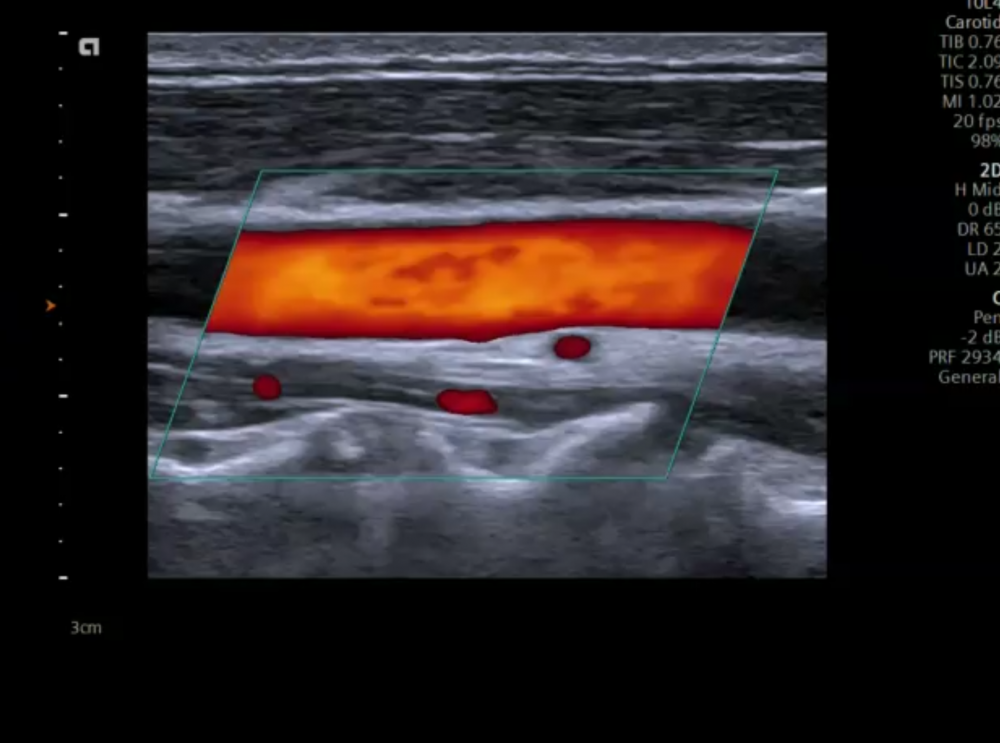
5. Color Doppler imaging of the carotid arteries
A) A brightness mode (b-mode) image of the lateral abdominal wall.
The A, B, M's – Ultrasound Modes Explained
Lensing B-modes in the Cosmic Microwave Background polarization
 Fable Fur Yarn in 15 Colors
 Lululemon Perfectly Overaized Crew Sweatshirt {Pink Savannah} 6 in 2023
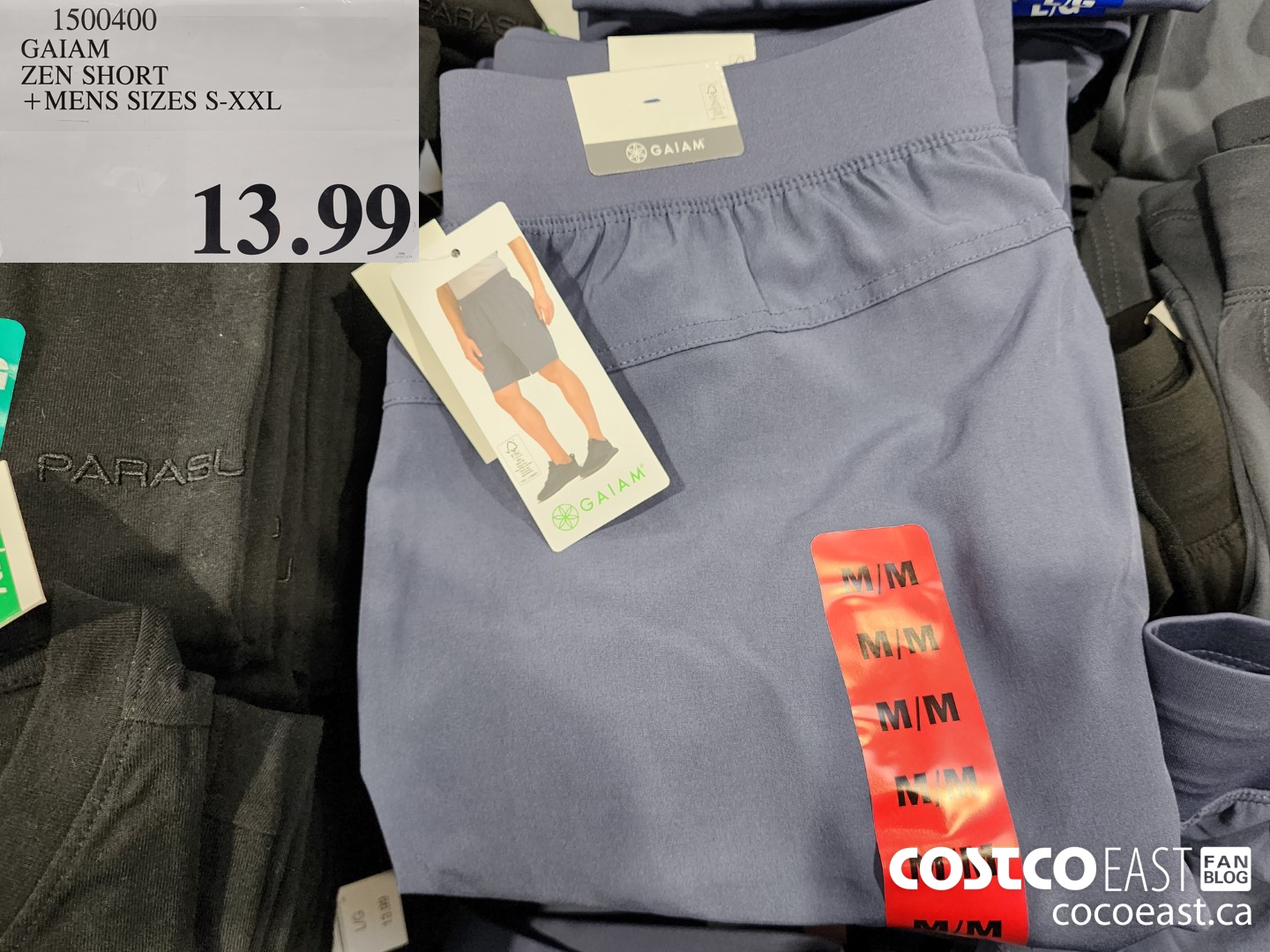 1500400 GAIAM ZEN SHORT MENS SIZES S XXL 13 99 - Costco East Fan Blog
 What is AMA? Understanding the Basics of Ask Me Anything
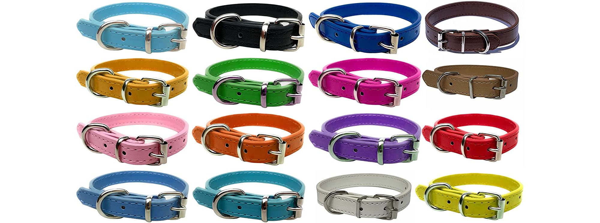 Strong Leather Dog Collar for Puppy, Cat, Dogs for Small, Medium, Large & Extra Large Pet Collars Range of Colours and Sizes
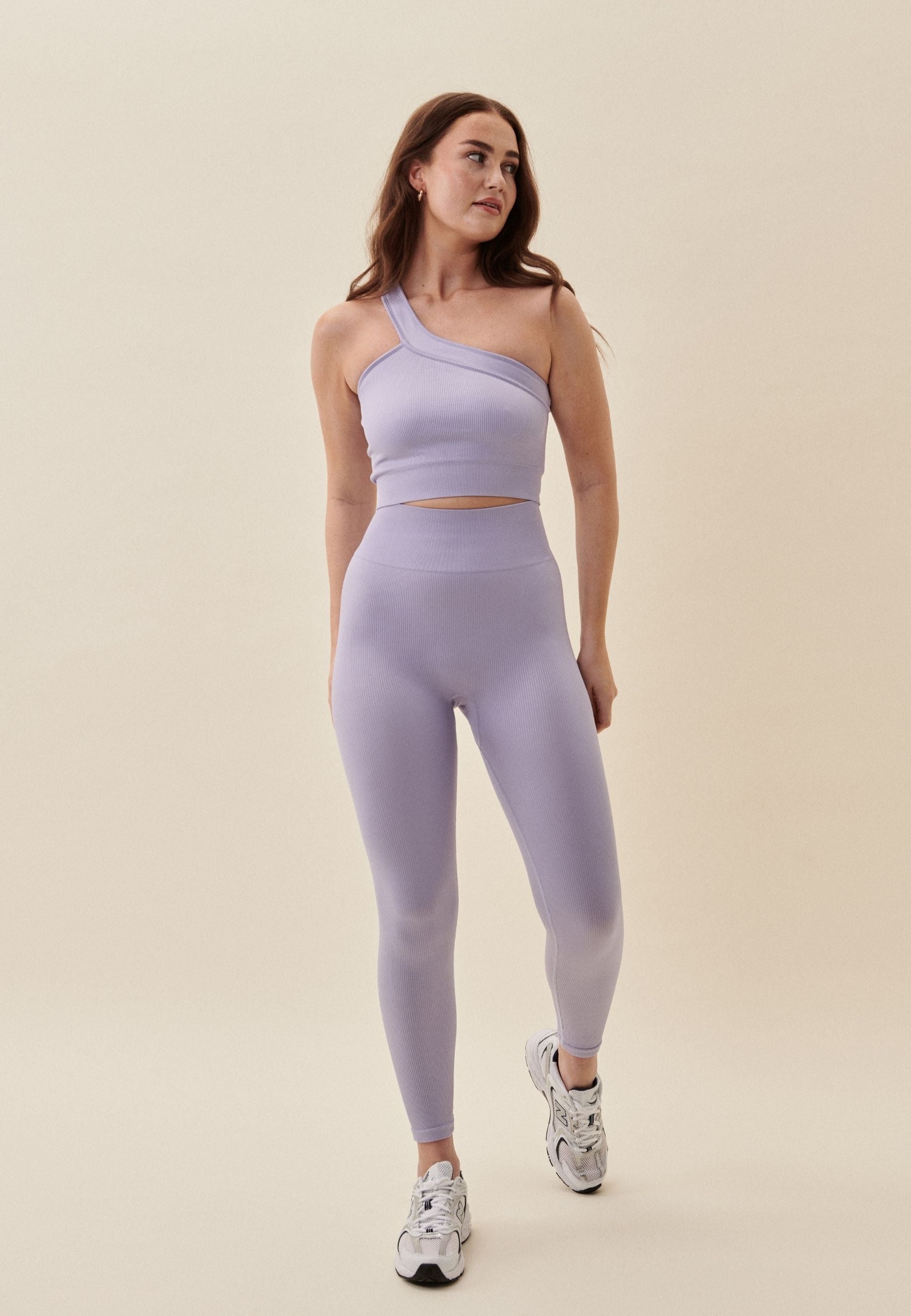 Ribbed Seamless Leggings - Lavender